One-pot formation and functional monitoring of neural cysts and organoids
Eindhoven University of Technology
THE NETHERLANDS
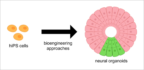
Three-dimensional (3D) cell spheroids and organoids have presented a new paradigm for studying the fundamental processes behind organ and tissue regeneration, as well as for developing treatments for diseases, in physiologically relevant 3D tissue contexts. However, robust and reproducible generation of spheroids and organoids remain challenging. In this talk, we will discuss several bioengineering approaches that we have developed to obtain neuroepithelial and neural organoids from human induced pluripotent stem cells in a controlled and tunable manner. The engineering-based approaches allow longitudinal monitoring and characterization, and are also shown to be particularly suitable for basic morphogenesis studies and disease modeling of neurodegeneration.
Novel single cell sequencing technologies
Singleron Biotechnologies
GERMANY

High throughput single cell sequencing is enabled by Singleron's cutting-edge technology which allows you to study genes and their functions across tens of thousands of cells simultaneously in a single experiment. It offers you essential insights into the samples with high level of heterogeneity and provide you with crucial information on gene expression across a multitude of cell subtypes present in your sample.The applications that go beyond gene expression profiling are gaining on popularity as well, as they enable you to obtain deeper insights on the function of specific genes by adding additional layers of information, such as for instance temporal resolution, to your data.
Development of a 3D cell model of skeletal muscle cells (myospheres) from human primary satellite cells.
University of Oslo
NORWAY
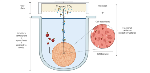
Nowadays, we know how muscle works in the body and how it can help us to counteract some diseases such as obesity. However, there is still a lack of information on how the muscle cells communicate with other organs in the body and how they respond or adapt to different situations at the molecular level. 2D cell models have been largely used to study energy metabolism concerning metabolic diseases but the result’s transferability to in vivo is limited. This project aimed to develop and characterize a human primary skeletal muscle 3D cell model (myospheres) as an easy, scaffold-free, and low-cost tool to later study impaired molecular mechanisms characteristic of metabolic disorders.
Non-invasive PET-imaging of CAR-T Cell Therapy using DTPA-R, a Novel Reporter Gene System
Technical University of Munich
GERMANY
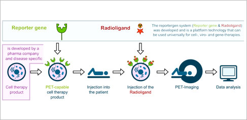
We developed a novel reporter gene system for non-invasive tracking of Cell-, Viro- and Gene-therapies using Positron Emission Tomography (PET), which consists of a membrane-anchored Anticalin binding protein (DTPA-R) and a corresponding radiohybrid radioligand [18F]F-DTPA. The simple design of the reporter protein with a minimal gene size yields high receptor densities of up to ~1x10⁶ receptors per T cell and highly specific binding of the corresponding radioligand [18F]F-DTPA was proven in preclinical proof-of-principle studies for CAR-T cell therapy. In this studies we quantified proliferation and migration of anti-CD19 CAR-T cells over one month of therapy.
New non-invasive, label-free monitoring approach for high cell culture and assay quality
Ludwig Maximilian University of Munich
GERMANY

Monitoring cell viability under culturing conditions provides a measure to address the principles of GCCP. Lensless Microscopy (LM) is ideal for this purpose since it allows cost-effective label-free live-cell imaging with compact instrumentation. We present a novel LM exploiting the optical properties of the cell itself as the imaging element or lens of the microscope. Cutting edge AI algorithms allow for fully automating the entire workflow from time-lapse imaging to proliferation and motility output parameters, leading to standardized protocols and constant conditions independent of the operator. We found, that heterogeneous cell culture conditions lead to an increase of variance during subsequent assays like omics-readouts. Enhancing the quality of cell culture ensures more reliable and reproducible results of subsequent experiments, like e.g. omics readouts, EC50 value determination, or treatment response. The approach of time-dependent multiparameter monitoring in real-time inside the incubator including the determination of key cell culture parameters including confluence, proliferation, and clustering as well as cell migration [3] further allows for standardization and hereby enhancement of the quality and reproducibility of different cell based assays, like wound healing assays, motility and proliferation assays, or spheroid growth monitoring. Continuous monitoring enhances the significance of toxicological and drug response assays compared to endpoint assays. Enabling faster and more reliable results of in vitro cell culture models, our approach contributes to the field of new approach methodologies in animal testing.
Automated manufacturing of 3D In Vitro test systems
Fraunhofer ISC
GERMANY
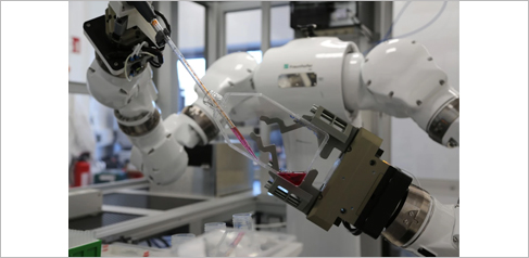
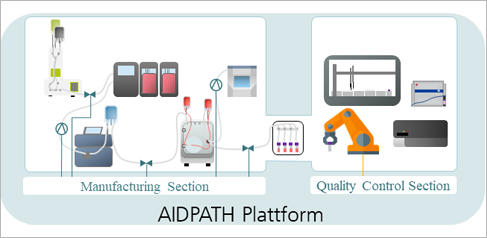
The Translational Centre for Regenerative Therapies (TLC-RT) of Fraunhofer ISC has a strong experience on the design, development and validation of human 3D tissue models with applications in basic and preclinical research. Our models are based on human primary cells, 3D organoid culture or different stem cell types such as human induced pluripotent stem cells (iPSCs) grown on suitable bioscaffolds. Recreation of innate physiological conditions is addressed by tissue culture in bioreactor platforms. Also, we are developing new technologies such as noninvasive assessment methods or automated tissue production to improve the quality and availability of the tissues. We will demonstrate two lead application in the automated manufacturing i.e. human 3D skin models as well as organoids.
Cellbox - the shipment solution for living cells, organoids and sensitive probes.
Cellbox Solutions
GERMANY

The trend to use complex human-like assay systems for drug discovery and drug profiling continues to increase. Also, therapeutic modalities like cells, tissues and mini organs are increasingly applied to patients. All these probes must not be frozen but need to be shipped to collaborators or patients at distinct conditions. The Cellbox offers a temperature, gas and pH controlled mobile incubator to ship such probes around the world. We will present the Cellbox in various application of such demand.
The modulation of cellular HCN4 channel trafficking in hiPSC-derived pacemaker-like cells during enteroviral infection with Coxsackievirus B3
WWU Münster
GERMANY
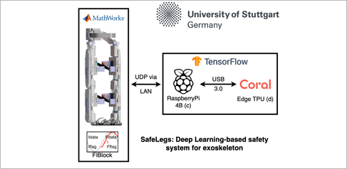
The SafeLegs is a supportive lower-limb 6-DOF exoskeleton demonstrator equipped with safety features. It is the result of collaboration between Uni Stuttgart and KIT. It consists of six actuators - brushless motors in the hips, knees, and ankles. Sensor signals are providing feedback from the motor encoders to the main system controller via the CAN bus. The SafeLegs is a complex Cyber-Physical System with components that prone to heterogeneous faults. To ensure safety we proposed a Deep Learning-based approach for system failure prevention. We employed LSTM network for error detection and mitigation. Trained LSTM models were quantized and deployed on an Edge TPU. The light weight of the system and minimal power consumption allowed its integration into wearable robotic systems.
A 3D cell culture taking into account the extracellular matrix for the development of liver organoid model.
HCS Pharmas
FRANCE
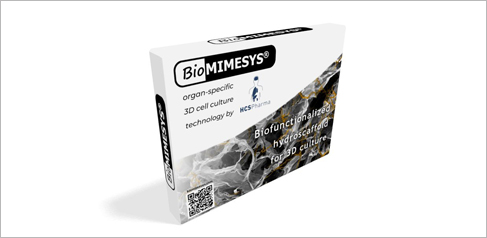
The extracellular matrix (ECM) is present in all tissues and is a master regulator of cellular behaviour and phenotype. We have developed 3D cellular models using BIOMIMESYS®, a patented hydroscaffold™ for 3D cell culture. In this presentation, we will exemplify the importance of the matricial micro-environment with the successful differentiation of human pluripotent stem cells (hiPSCs) into mature and functional liver cells within BIOMIMESYS®. This model provides a suitable model to study healthy liver functions but also metabolic diseases upon hepatocyte-like cell (HLC) differentiation. Our 3D in vitro models mimicking the ECM microenvironment should help to model in vivo complexity with more relevance, and therefore to discover new effective therapies against diseases like cardiometabolic conditions.
Hetero-spheroids for the study of the cross-talk between tumor cells and CAFs in different metastatic cancer models
University of Milan-Bicocca - Department of Biotechnology and Biosciences
ITALY

Desmoplastic tumors are highly aggressive and difficult to eradicate due to the complex interactions between epithelial cells and microenvironment, which play a key role in stimulating pro-survival and progression programs. The cross-talk between cancer-associated fibroblasts (CAFs) and tumor cells, in particular, is responsible for the high invasivity of these tumors and resistance to chemotherapy. Hetero-spheroids obtained by co-culturing fibroblasts and epithelial cells of pancreatic and metastatic breast cancer were developed to determine the molecular and functional changes obtained upon different cell types interaction. The role of some soluble mediators (TGFβ and IL-6) in the cross-talk CAFs-tumor cells has been investigated. Identification of involved mediators and pathways is of great importance for the identification of new therapies against these malignant tumors.
